








ISTANBUL EYE HOSPITAL
Our hospital has been serving as a private branch hospital since 2001.
Istanbul Eye Hospital, which provides service in Bahçelievler, has transportation connections with districts such as Yeşilyurt, Florya, Ataköy, Bakırköy, Şirinevler, Bagcilar, Güngören, Avcılar. Incirli Metrobus Stop is 500 meters away and Ataturk Airport is 7 km away.
To provide eye disease diagnosis and treatment services by ad homing employee and patient rights by ad hosing medical ethics and work ethic.

SERVICES & SERVICES
GENERAL EYE EXAMINATION
EXCİMER LASER
Cataract
Glaucoma
Slt
SQUINTY AND CHILD’S EYE
Retina
Uveitis
AMD (Age-Related Mathesia
RELIGION AND EYE
CORNEA AND DISEASES
KERATOCONUS AND TREATMENT
NEURO-OPHTHALMOLOGY
OPHTHALMIC PLASTIC SURGEON
TEARWAYS DISEASES
EYE TUMORS
COLOR BLINDNESS (DALTONISM)
CONTACT LENS
ADVANCED TREATMENTS AND TREATMENTS
FREQUENT EYE PROBLEMS
Astigmatism:
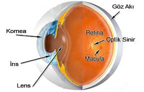
It’s one of the re offending defects. It affects the focus of light on the retina, which impairs the interpretation of the image in the brain. Astigmatism is usually caused by the fact that the oblique of the cornea has a light egg shape. It is not a disease and is a very common problem. There is blurred vision in both distant and close objects, which causes eye fatigue, eye slippage and disturbedness in people. Sometimes astigmatism is with myopia and hyperopia.
These disorders, called myopic astigmatism and Hyperopia astigmatism, can be treated with glasses and contact lenses.
Astigmatism
Myopia:
The person sees nearby objects blurred in the clear away, and the front rear length of the eye is more than normal or the cornea’s course is too steep. The image (Images) focuses in front of the yellow dot. Myopia begins under the age of 20, usually at the age of 8-12 years. It can progress rapidly in adolescence. Correction is made with glasses and contact lenses.
Myopia
Hyperopia:
Remote objects are seen blurred in the net nearby. After close work, blurred vision, discomfort, fatigue occur in the person. Hidden hyperopia can be overlooked in sloppy examinations. The eyeball in the hyperopia eyeball is small or the cornea is very flat. The image focuses on the back of the eye.
Hyperopia
Cataract:
It is a loss of transparency that develops in the eye’s own lens. It causes foggy-blurred vision. The incidence increases over the age of 55. Alhowever, there is no definitive way to prevent ultraviolet rays and smoking are thought to accelerate the development of catalalyt. Catalyte significantly impairs vision in late stages.
Cover Oil Kit:
It is the formation of swelling in the eyelids that can be mixed with shallots. It is an inflammation of the small fat-producing gy gy gies in the upper and lower eyelids. This occurs by clogging the g g g gies. It can shrink and disappear on its own, as well as hot compresses can inject drugs into swelling and sometimes surgery may be required. Frequent recurrent shalasions can be symptoms of other diseases.
Conjunctivitis:
It is inflammation of the thin membrane that covers the white part of the eye. Bacterial infections, viral infections, allergies, environmental pollution (smoke and chemical vapor) can lead to this condition. With the increased blood flow in the conjuncture, the eye appears red. It can be acute (intense), chronic (long-term). There may be burring and adhesions in the eyelashes in the morning.
Allergic Conjunctivitis:
It is common in people with hay flu or seasonal allergy. Usually in the spring the symptoms increase. Red itchy eyes and watering happens. Animal feathers, dust, pollen are the most common causes. Although it is easy to treat, sometimes it can be annoying.
Dry Eye:

Tears are very important for eye health. It contains natural lubricant and preservatives of the eye. People with constant eye sanks may have dry eyes. The symptom of the dry eye can sometimes be paradoxic watering. This occurs reflexively against stings. Causes of dry eyes; Decreased blink reflex, antihistamine drugs, environmental factors (low dehydrta and wind), chemical and thermal burns and some health problems such as arthritis. Sucterial tear drops, gels, sometimes an unspothed blockage of the tear ducts are used for treatment purposes. Room humidifiers can be useful in dry areas.
Glaucoma (Eye Pressure):
2 out of every 100 people over the age of 35 have glaucoma. The risk is more in people with glaucoma or myopia in their families. Early diagnosis is very important with regular eye examinations, as the disease cannot be prevented and the damage caused to the eye nerve cannot be reversed. There are different types of glaucoma. But they all ultimately damage the eye nerve. After the eye’s own liquid is produced, it leaves the eye in the sponge membrane between the cornea and sclera. A blockage in this area causes eye pressure to rise. Symptoms vary depending on the type of glaucoma.
Chronic Open Angle Glaucoma:
He’s the most common type. He’s over 40 years old and symptomless, so he’s silent. It usually causes im im im possible damage to the patient before diagnosis. That’s why regular eye pressure measurement is so important.
Normotensive Glaucoma:
Although intraocular pressure is possible at normal limits, glaucoma is due to impaired nutrition of the eye nerve (optic disc). In people with thin corneas, the actual value of intra-eye pressure in the eye is more than obtained. In this case, the eye nerve is affected. In addition, normotensive glaucoma occurs in some people (elderly) with normal eye pressure due to lack of blood intake of the eye nerve.
Birth glaucoma:
It appears immediately after birth. There is a problem with the evacuation system of eye fluid. Babies with sensitivity to light or constantly watering eyes should be examined especially for this condition.
Acute Angle Closure Glaucoma:
Eye fluid is caused by the sudden and complete closure of the evacuation area. The patient sees halo (ring) around the lights, there is severe pain, nausea and blurred vision. He could be any age. Emergency treatment is required. Blindness may develop within 1-2 days.
Secondary Glaucoma:

It is glaucoma caused by another eye problem. Like uve-related glaucoma.
Maestro Degeneration:
The ma’an is located at the center of the retinal layer and is responsible for central vision. In this disease, central vision is disturbed. There are two types. Dry type and age type. The incidence increases over the age of 50. That’s why it’s called age-related maula degeneration.
Dry Type – Maethy Degeneration:
That’s 90%. Patients see a black mark in the center or describe a break in thin long objects. It shows slow progress over the years.
Age Type – Maethy Degeneration:
There is a new formation of vessels in the machine, which tends to bleed easily and leak fluids. Central vision can be quickly lost. With examinations such as FFA/ICG, the location of new vessels can be determined and control can be achieved with Argon Laser or photodynamic treatment.
FlyFly Flights:
Sometimes people can see specks and threaded objects in the area they are looking at. Although they can be seen at any age, they are more frequent at older ages. A gel-like substance normally called vitreus fills the eye. The remains of the birthal protein in this substance, water cavities can be monitored as spots in the field of vision. Fly-flying can often indicate an eye disease, where it can be a normal condition. (Uveitis, bleeding in the eye, retinal tear, etc.)
Ail:
It is caused by the formation of red swelling in the lids. Eyelash is inflammation of the follicle. Hot compresses and antibiotic pomads are used in the treatment.
Blepharitis:
It is chronic and stubborn eyelid inflammation that is common in people with oily skin, dandruff, dry eyes. There is discomfort in the eyes, itching, dandruff at the bottom of the eyelashes and slight redness. Alhowever, there is no definitive treatment, control can be achieved with the medications given by the eye doctor.
Back
VISUAL IMPAIRMENTS AND TREATMENT
Normal Eye
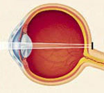
If the rays broken in the cornea and lens fall exactly on the retina into the visual center, there are no resection defects and a clear view is obtained.
Myopia (Not seeing far)
While the remns should focus on the retina, they are focused in front of the retina. With the rays broken in the cornea and lens focusing clearly on the retina before reaching the center of vision, the eye cannot see the objects in the away area clearly. It’s called Myopia.
Hyperopia (Insurmology)
The remns can’t be broken enough to focus on the retina. The eye relatives cannot see clearly as the rays broken in the cornea and lens focus more further on the retina, not on the visual center. This condition is defined as Hyperopia.
Astigmatism
The remns focus more than a point in the retina. For a clear view, the cornea must be smooth and in the same curve on each axis. More or less curved cornea on a certain axis reveals astigmatism defect. The image is not clear far away or nearby, and the person sees objects shaded.


